Why are doctors not looking for Kaposi's Sarcoma in Chronic Fatigue Syndrome patients?
Biopsy
To be sure that a lesion is caused by KS, the doctor will need to take a small sample of tissue from the lesion and send it to a lab to be checked. This is called a biopsy. A specially trained doctor called a pathologist can often diagnose KS by looking at the cells in the biopsy sample in the lab.
For skin lesions, the doctor will usually perform a punch biopsy, which removes a tiny round piece of tissue. If the entire lesion is removed, it is called an excisional biopsy. These procedures can often be done with just local anesthesia (numbing medicine).
Lesions in other areas, such as the lungs or intestines, can be biopsied during other procedures such as bronchoscopy or endoscopy, which are described below. Since biopsy of lesions in these areas can sometimes cause serious bleeding, biopsy is often not done in people already known to have KS.
https://www.cancer.org/cancer/kaposi-sarcoma/detection-diagnosis-staging/how-diagnosed.html
This is what oral Kaposi's Sarcoma looks like.
Source: https://doctorspiller.com/kaposis-sarcoma/
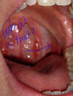
Compare it to these crimson crescent lesions in the mouths of Chronic Fatigue Syndrome patients.
"Burke A. Cunha, MD, discovered what he called crimson crescents in the mouths of 80% of his CFS patients. After the word got out, Cunha received calls from other parts of the country. Physicians began telling him that they also were finding the crimson crescents in their patients once they looked for them."
https://www.prohealth.com/library/crimson-crescents-facilitate-chronic-fatigue-syndrome-cfs-diagnosis-11266
Chronic Fatigue Syndrome patients may have undiagnosed internal Kaposi's Sarcoma. Susan Levin found HHV-8, the Kaposi's Sarcoma virus, in half of CFS patients she looked at.
Prevalence in the Cerebrospinal Fluid of the Following Infectious Agents in a Cohort of 12 CFS Subjects
Susan Levine
Published online: 04 Dec 2011
ABSTRACT
Over the last decade a wide variety of infectious agents has been associated with the chronic fatigue syndrome (CFS) as potential etiologies for this disorder by researchers from all over the world. Many of these agents are neurotrophic and have been linked previously to other diseases involving the central nervous system (CNS). Human herpes virus-6 (HHV-6), especially the B variant, has been found in autopsy specimens of patients who suffered from multiple sclerosis. Because patients with CFS manifest a wide range of symptoms involving the CNS as shown by abnormalities on brain MRIs, SPECT scans of the brain and results of tilt table testing we sought to determine the prevalence of HHV-6, HHV-8, Epstein-Barr virus (EBV), cytomegalovirus (CMV), Mycoplasma species, Chlamydia species, and Coxsackie virus in the spinal fluid of a group of 12 patients with CFS. Although we intended to search mainly for evidence of actively replicating HHV-6, a virus that has been associated by several researchers with this disorder, we found evidence of HHV-8, Chlamydia species, CMV and Coxsackie virus in 6/12 samples. Attempts were made to correlate the clinical presentations of each of these patients, especially the neurological exams and results of objective testing of the CNS, with the particular infectious agent isolated. It was also surprising to obtain such a relatively high yield of infectious agents on cell free specimens of spinal fluid that had not been centrifuged. Future research in spinal fluid analysis, in addition to testing tissue samples by polymerase chain reaction (PCR) and other direct viral isolation techniques will be important in characterizing subpopulations of CFS patients, especially those with involvement of the CNS.
https://www.tandfonline.com/doi/abs/10.1300/J092v09n01_05


